Thursday, October 27, 2011

Selective & Differential Results

Taking our selective & differential agar plates, we observed the EMB, Mannitol and MacConkey results for bacteria 'L'. The Mannitol plate was negitive, this means our bacteria did not grow and does not tolerate salt and cannot ferment Mannitol. This means our bacteria is not a halophile (therefore not of the staphylococcus genus). Our bacteria did grow on the MacConkey agar & was pinkish colored on the EMB agar! This means our bacteria is labeled as gram-negaticve enteric bacteria and partially ferments lactose.

Mannitol

EMB
Hydrogen Peroxide was also used in class & placed onto of the bacteria to test for the enzyme catalyse. As the hydrogen peroxide touched bacteria 'L' bubbles formed meaning that the bacteria successfully broke down the hydrogen peroxide into water and that it is an aerobe (which we knew since ours is facultative) and does contain the enzyme catalyse!!
Identity of 'L' is almost clear!!
Later,
Annie B

Tuesday, October 25, 2011

Litmus Test Results & Mannitol, EMB, and MacConkey Tests

Our litmus milk broth turned a slightly pinkish but still mostly purple blue. We think it is slightly acidic. And there are curds in the broth. We also preped for several selective and differential tests today, sepecifically the Mannitol, EMB (Eosin Methylene Blue Agar) and MacConkey Tests. Using aseptic technique, we inoculated bacteria 'L' onto these three different Agars. They were then placed in the incubator at 25 degree Celsius. They will be checked next week to determine which of the agars bacteria 'L' thrives on!!

Anne B

Thursday, October 20, 2011

Five Tests & Four Results [ONE TO GO]

It's here! The results from four of the five tests from last lab! [Remember litmus milk test takes a week to get results from].
First up is the Gelatin Hydrolysis Test. After placing the same in the refrigerator for 15 minutes we checked the gelatin tube for liquidation. Then we compared it to the control tube we placed in (of just the gelatin). Result: POSITIVE! Our tube did in fact have liquid!! This means that there is gelatinase in bacteria 'L' because it consumed the gelatin!
Test 2 was the fermentationi of carbohydrates! Upon observation it was determined that our bacteria ferments glucose and sucrose but it does not ferment lactose. Test 3 was TSIA and bacteria 'L' had a red slant and yellow butt of slant. There was also a gas bubble that split the butt! (acidic and produces CO2). This means only glucose was fermented. Last but not least was the results for teh MR-VP! Once we pulled the tube out of the incubator we slit it into two more tubes. In one tube we added 5 drops of Methyl Red de and in the other tube we added 5 drops of VP-B and 15 drops of  VP-A. Our bacteria was negative fore the Methyl Red Test but Positive for the Voges-Proskauer due to a red band identified near the top of the tube. This means it ferments by Butanediol Fermentation and breaks acids down into non-acids!

Gelatin Hydrolysis Liquidation
Lactose

TSIA (notice the air bubble on the right side of butt)

Sucrose

VP-positive (left) MR-negitive (right)
Glucose & Sucrose


Annie B

Tuesday, October 18, 2011

Putting 'L' To The Test (Well actually to many tests...)

After an amazing field trip to the local water filtration plant last Thursday, we are back at 'L' in the laboratory. We addressed five tests: Gelatin Hydrolysis, Fermentation of Carbohydrates, Methyl-Red/Voges-Proskauer (MR-VP), Triple Sugar Iron Agar (TSIA) and a litmus milk test. Today, tuesday, we prepared for all these tests and will check the results next lab! (except the litmus test which takes a week to incubate so that will be checked than!)
First up, we have the Gelatin Hydrolysis Test! To prepare for this test we used the aseptic technique with a inoculating needle to stab our bacteria into a gelatin agar slant. We made sure the bacteria made it into the gelatin agar slant by stabbing the needle 3/4 of the way through the gel! Then the slant was placed into teh incubator at 25 degree Celsius. If the bacteria digests the gelatin, than a liquid should appear in the tube when we check it! If it does indeed digest gelatin, then our bacteria has gelatinase, the enzyme that digests gelatin.

The 2nd test was the fermentation of carbohydrates. This test used a broth tube that contained phenol red-sugar broth & a durham tube (which traps gas bubbles). There were three of these tubes and each contained a different sugar. There was a sucrose, a lactose, and a glucose broth. Using aseotic technique we placed our bacteria into each tube to see which carbohydrates our bacteria can digest. We placed the tubes into the incubator at 25 degrees Celsius.

The next test was the Methyl Red/ Voges-Proskauer (MR-VP) test. We used an MR-VP broth and (using aseptic technique) we put bacteria 'L' into the broth tube. Then the broth was placed in the incubator at 25 degree Celsius. Fourth was the Triple Sugar Iron Agar (TSIA) test. We used the aseptic technique & a inoculating needle to transfer bacteria 'L' 3/4 of the way into a TSIA slant. This too was placed in the incubator at 25 degrees Celsus. The last test was the litmus milk test. Using aseptic technique (again! it never gets old ^_^) bacteria 'L' was placed into a Litmus Milk based broth and then into the incubator at 25 degrees Celsius.


Next class we will see the results!
Annie B

Tuesday, October 11, 2011

GasPak Results & Food Source of Bacteria 'L'

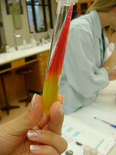
Our bacteria 'L' did infact thrive in the GasPak from last week. This means 'L' can grow in both aerobic & anaerobic conditions. This means our hypothesis from earlier was correct and our bacteria is facultative (it can grow in the presence & absence of oxygen). The Thioglycollate Broth also showed the same result: that 'L' is facultative. 'L' grew throughout the broth: present equally in both the top, red layer (where oxygen is present) and in the bottom yellow layer (where oxygen is not present).
Once we determined the oxygen requirements, we did several tests to figure out what 'L' ate. We took three nutrient agar plates each with a different nutrient. One contained starch, another had casein (a milk protein), and the thrid contained triglycerides (lipids). Using the aseptic technique & a inoculating loop, we spread bacteria 'L' onto all three plates and incubated them at 25 degree Celcius overnight.
We took the three out and examined the results. We had to cover the Starch agar plate with Gram's Iodine for 1 minute to see if the enzyme Amylase was present in our Bacteria. If it was a halo would appear around our bacteria streak. The casein plate was to see if the enzyme Casein Protease was present in our Bacteria. As shown by the halo around the bacteria, Bacteria 'L' does indeed contain Casein Protease! The triglyceride plate was to see if the enzyme Lipase was present in our bacteria. The dark blue color in our sample proves that Bacteria 'L' does eat triglycerides and contains Lipases.
Starch Agar with Gram's Iodine (negitive test)

Casein Agar (positive test)

Triglyceride Agar (positive test)

An upclose look at the halo atop Bacteria 'L'
Peace,
Annie B

Thursday, October 6, 2011

Is 'L' Anaerobic? & Storing Bacteria Forever

Today in lab we discovered how to store bacteria forever! By using Glycerol to slow down the rapid cooling of the bacteria membrnes from the refrigerator (this causes the bacteria membranes to burst). We first used 300 microLiters of Glycerol (100%) using a micropipet. We than placed this into a small storing capsule and replaced the plastic covering on the micropipette to prevent contamination of the bacteria. Than with the new tip we measured 700microLitters of the bacteria in broth and added it to the 100% Glycerol and mixed them together. Once properly labeled we placed the capsule in a -80 degree Celsius refridgertor on the rack that read 'FOR NURSING MAJORS ONLY' so it didn't get confused with Biology majors samples!
We than used Thioglycollate Broth to determine if 'L' was anaerobic or not! This test is positive if the Thioglycollate does not turn red (red is where the chemical reacted with oxygen). Using the aseptic technique, we placed 'L' into the broth and left it in a 25 degree incubator. If the bacteria grows in the yellow portion of the test tube it is anaerobic and if it goes in the red it is aerobic.

Micropipetting The 100% Glycerol

Our bacteria that is going to live FOREVER!

GasPak & Thioglycollate Broth


As a class, we used a GasPak to see if our bacteria was anaerobic. Our sample was placed on a nutrient agar plate (using aseptic technique & inoculating loop) and placed inside the Pak. The Pak eliminates oxygen (as seen on the colorless strip that appears blue when oxygen it present!) Instead CO2 is inside the Pak. If our bacteria 'L' does grow it means that it is anaerobic. Since it has already been thriving in aerobic conditions it really means that it is facultative.

Until Next Lab,
Annie B

Tuesday, October 4, 2011

Acid-fast & Endospore Stains

This week started off with an endospore stain. We first begin a simple stain (smearing, air-drying & heat fixing-mentioned in previous posts!) and than placed the slide in a slide drying rack suspended over a beaker filled with boiling water. After covering the smear with Bibulous paper we wet the paper with Malachite Green dye. We continued to add dye to the paper, keeping it damp for around 5 minutes. We did not let all of the stain evaporate, therefore the slide could properly set but not kill the bacteria! After 5 minutes, we removed the slide with forceps (twisers) and set the slide on paper towels to cool down. The slide was than rinsed with distilled water and remove some excess Malachite Green from the slide. Then we added Safranin dye for 60 to 90 seconds over a sink on a drying rack. Once correct time had passed, we rinsed with distilled water and let dry. Once viewed under a microscope it was apparent that the Green Malachite dye had all rinsed away and only the Safranin stained bacteria remained. This meant that bacteria 'L' did not hace endospores as the Green Malachite dye reminds when there are endospores.

Next we did an acid-fast stain. We started with a simple stain and than suspended it over boiling water on a drying rack. Once covered with Bibulous paper, we saturated it with Ziehl-Neelsen Carbolfuchsin. We continued to keep the slide wet with dye for 3 to 5 minutes. Once time was up, we used forceps to remove the slide to cool down and to throw away the Bibulous paper into the proper waste. The slide was then rinsed with distilled water to remove excess dye and to remove even more stain we took acid-alcohol and dropped it over the slide, which we placed at a 45 degree angle over the sink, until the pink color of the Ziehl-Carbolfuchsin was done running off. After once again using distilled water to rinse, we made sure to cover the smear with Methylene Blue for 2 minutes, rinsed it clean with more distilled water and than blotted dry with Bibulous paper.
Since bacteria 'L' remained dark pink after using acid-alcohol it is determined that our bacteria is acid-fast! This means the dark pink dye was trapped by the waxes in the cell membrane of bacteria 'L'!
Methylene Blue rinsing off!           
Annie B